The simplest unicellular organisms do not have nervous system, regulation of vital activity in them occurs only due to humoral mechanisms. At the same time, under the influence of any factor of the external or internal environment, the production of regulatory molecules increases, which are released directly into the intracellular fluid (lat. " humor"- liquid) and enter the working organelle by diffusion. Only after this is formed a response to the causative factor. Of course, this method of regulation is not operational and accurate, and, therefore, limits the adaptive capabilities of the organism.
The nervous system, which appeared in multicellular organisms, allows you to control the body systems more differentiated and with less time lost to conduct a command signal (stimulus). Therefore, in all modern highly organized animals, with a single neurohumoral regulation of body functions, the leading role belongs to the nervous system. Phylogeny of the nervous system, that is, its evolutionary development (Greek “ phylon"- genus), presumably, took place in several stages:
Stage I - the formation of the network nervous system. At the present stage of evolution, coelenterates, for example, hydra, have this type of nervous system (Fig. 12). All their neurons are multipolar and unite due to their processes into a single network that permeates the entire body. When any point of the hydra's body is irritated, the entire nervous system is excited, causing the movement of the whole body. The evolutionary echo of this stage in humans is the network-like structure of the intramural * nervous system of the digestive tract (metasimpathetic autonomic nervous system).
Stage II - the formation of the nodal nervous system associated with the further integration of the organism and the need for centralized processing of information to speed up this process. At this stage, there was a specialization of neurons and their convergence with the formation of nerve nodes - centers. The processes of these neurons formed nerves leading to the working organs. The centralization of the nervous system led to the formation of reflex arcs. The process of centralization took place in two ways (Fig. 13): with the formation of a radial (asymmetric) nervous system (echinoderms, mollusks) and a ladder (symmetrical) system (for example, flat and roundworms).
The radial nervous system, in which all the nerve ganglia are concentrated in one or two or three places, turned out to be of little evolutionary promise. Of the animals with an asymmetric central nervous system, only octopuses have reached the lowest level of the perceptual psyche, while the rest have not risen above the sensory psyche.
During the formation of the ladder-type CNS (as, for example, in planarians, see Fig. 13, A), ganglia form in each segment of the body and are connected to each other, as well as to segments of the upper and lower levels through longitudinal trunks. At the anterior end of the nervous system, ganglions develop that are responsible for receiving information from the anterior part of the body, which, in the process of movement, first and more often encounters new stimuli. In this regard, the head ganglia of invertebrates are more developed than others, being the prototype of the future brain. A reflection of this stage in the formation of the central nervous system in humans is the structure of the autonomic nervous system in the form of parallel chains of sympathetic ganglia.
Stage III is the formation of the tubular nervous system. Such a central nervous system first arose in chordates (lancelet) in the form of a metameric * neural tube with segmental nerves extending from it to all segments of the body - trunk brain(Fig. 14). The appearance of the trunk brain is associated with the complication and improvement of movements that require the coordinated participation of muscle groups of different segments of the body.
Stage IV is associated with the formation of the brain. This process is called cephalization(from the Greek. " encephalon" - brain). Further evolution of the CNS is associated with the isolation of the anterior part of the neural tube, which was initially due to the development of analyzers, and adaptation to various environmental conditions (Fig. 15).
Phylogeny of the brain, according to the scheme of E.K. Seppa et al. (1950), also goes through several stages. At the first stage of cephalization from the anterior part of the neural tube three primary bubbles. Development posterior bladder(primary rear, or rhomboid brain, rhombencephalon) occurs at lower fish in connection with the improvement of the auditory and vestibular analyzers that perceive sound and the position of the body in space (VIII pair of head nerves). These two types of analyzers are most important for orientation in the aquatic environment and are probably the earliest in evolution. Since the hindbrain is the most developed at this stage of evolution, the control centers of plant life are also laid in it, which control critical systems the life support of the body - the respiratory, digestive and circulatory systems. Such localization is also preserved in a person in whom the above centers are located in the medulla oblongata.
The hindbrain is divided into hindbrain proper (metencephalon), consisting of the pons and cerebellum, and medulla (myelencephalon), which is transitional between the brain and spinal cord.
At the second stage of cephalization there has been a development second primary bubble (mesencephalon) under the influence of the visual analyzer that is being formed here; this stage also began in fish.
In the third stage of cephalization formed forebrain (prosencephalon), which first appeared in amphibians and reptiles. This was due to the exit of animals from the aquatic environment into the air and the enhanced development of the olfactory analyzer, which is necessary to detect prey and predators at a distance. Subsequently, the forebrain divided into intermediate and telencephalon (diencephalon and telencephalon). The thalamus began to integrate and coordinate the sensory functions of the body, the basal ganglia of the telencephalon began to be responsible for automatisms and instincts, and the cortex of the telencephalon, which was originally formed as part of the olfactory analyzer, eventually became the highest integrative center that forms behavior based on acquired experience. The issues of telencephalon evolution will be discussed in more detail in Section 6.5.1.
Stage V of the evolution of the nervous system - corticolization of functions(from lat. " cortex"- bark) (Fig. 16). The cerebral hemispheres, which arose in fish as paired lateral outgrowths of the forebrain, initially performed only an olfactory function. The cortex formed at this stage and performing the function of processing olfactory information is called ancient bark (paleocortex, paleocortex). It is distinguished by a small number of layers of neurons (2-3), which is a sign of its primitiveness. In the process of further development of other parts of the cerebral cortex, the ancient cortex shifted downward and medially. At different types it retained its function, but its relative dimensions decreased. In humans, the ancient cortex is present on the inferomedial surface of the temporal lobe (the anterior perforated substance and adjacent areas), and is functionally part of the limbic system and is responsible for instinctive responses (see Section 6.5.2.1.2.).
Starting with amphibians (see Fig. 16), the formation of the basal ganglia (structures of the striatum) and the so-called old bark (archicortex, archicortex) and their importance in the formation of behavior increases. The basal ganglia began to perform the same function as the archicortex, greatly expanding the range and complexity of automatic, instinctive responses.
The old cortex, like the ancient one, consists of only 2-3 layers of neurons. In amphibians and reptiles, it occupies the upper parts of the cerebral hemispheres. However, beginning with primitive mammals, as the new cortex enlarges, it gradually shifts to the median surface of the hemispheres. In humans, this type of cortex is located in the dentate gyrus and hippocampus.
The old cortex is included in the limbic system, which also includes the thalamus, amygdala, striatum, and ancient cortex (see Section 6.5.2.1.2.).
With the formation of this system, the brain acquires new functions - the formation of emotions and the ability for primitive learning based on positive or negative reinforcement of actions. Emotions and associative learning have greatly complicated the behavior of mammals and expanded their adaptive capabilities.
Further improvement of complex forms of behavior is associated with the formation of a new cortex ( neocortex, neocortex). Neurons of the neocortex first appear in higher reptiles, however, the neocortex is most developed in mammals (see Fig. 16). In higher mammals, the neocortex covers the enlarged cerebral hemispheres, pushing down and medially the structures of the ancient and old cortex. The new cortex is most developed in humans, its area reaches 220,000 mm 2, while two-thirds of the cortex area is in its folds. The neocortex becomes the center of learning, memory and intelligence, can control the functions of other parts of the brain, influencing the implementation of emotional and instinctive forms of behavior.
Thus, the significance of corticolization of functions lies in the fact that, as it develops, the cortex of the telencephalon takes on the role of a higher center for processing information and building programs of behavior. At the same time, the cortical sections of the analyzers and the cortical motor centers subdue the underlying evolutionarily older centers. As a result, information processing is improved, since qualitatively new capabilities of the cortex are added to the integrative capabilities of the subcortical centers. Phylogenetically old sensory centers become switching centers, carrying out the initial processing of information, the final assessment of which will be made only in the cerebral cortex.
The formation of behavior is built according to the same scheme: instinctive, species-specific automatic actions are regulated by subcortical nuclei, and acquired components of behavior that are developed throughout life are formed by the cortex. The bark, on the other hand, can control the centers of instinctive reactions, while significantly expanding the range of behavioral reactions.
Corticolization of functions increases with the transition to more high level evolutionary development and is accompanied by an increase in the area of the crust and an increase in its folding.
Reticulated. It first occurs in multicellular animals - coelenterates.
Heavy. Characteristic of lower worms.
Nodal. Characteristic of annelids and arthropods.
tubular. characteristic of chordates.
4. 1. Reticulate, diffuse nervous system . It arises in freshwater hydra, has the shape of a grid, which is formed by the connection of process cells and is evenly distributed throughout the body, thickening around the oral appendages. The cells that make up this network differ significantly from the nerve cells of higher animals: they are small in size, do not have a nucleus and a chromatophilic substance characteristic of a nerve cell. This nervous system conducts excitations diffusely, in all directions, providing global reflex reactions.
2. Heavy. In lower worms. Nerve cells are not scattered throughout the body, like in hydra, but are collected in two nerve trunks. In the front part they thicken - a paired head ganglion, which begins to play a leading role.
2. Nodal. In annelids and arthropods. The main achievement is segmentation, chains of nerve nodes are formed that “serve” certain parts of the body. Enlargement of the size of the head.
3. tubular the nervous system (in vertebrates) differs from the nervous system of the worm-like ones in that skeletal motor apparatuses with striated muscles arose in vertebrates. This led to the development of the central nervous system, the individual parts and structures of which are formed in the process of evolution gradually and in a certain sequence. cranium.
Centralization is a process of accumulation of nerve cells, in which individual nerve cells and their ensembles began to perform specific regulatory functions in the center and formed the central nerve nodes.
cephalization- this is the process of development of the anterior end of the neural tube and the formation of the brain, associated with the fact that nerve cells and endings began to specialize in receiving external stimuli and recognizing environmental factors. Nerve impulses from external stimuli and environmental influences were promptly transmitted to the nerve nodes and centers.
In the process of self-development, the nervous system consistently goes through critical stages of complication and differentiation, both in morphological and functional terms. The general trend of brain evolution in ontogenesis and phylogenesis is carried out according to a universal scheme: from diffuse, weakly differentiated forms of activity to more specialized, local forms of functioning.
6. Depending on the structural and functional features of the innervated organs, somatic and vegetative parts of the nervous system. Somatic nervous system - part of the nervous system that regulates the activity of the skeletal (voluntary) muscles. Vegetative nervous system - part of the nervous system that regulates the activity of smooth (involuntary) muscles of internal organs, blood vessels, skin, heart muscles and glands. In turn, depending on the anatomical and functional features, the autonomic nervous system is divided into two sections: sympathetic and parasympathetic.
Somatic department The nervous system is represented by cranial and spinal nerves.
Vegetative department The nervous system is represented by parasympathetic, sympathetic and metasympathetic innervation, each of which has a number of features.
The autonomic nervous system consists of autonomic neurons located in the middle, medulla and spinal cord, as well as in the ganglia on the periphery. It is characterized by a two-neuron principle of education.
The central part of the autonomic nervous system consists of the first neurons located in the middle, medulla oblongata and spinal cord.
The peripheral link of parasympathetic and sympathetic innervation is a chain of two series-connected neurons. The axons of the first neurons leave the CNS and necessarily end on the second neurons, united in the ganglion. The axons of the second neurons go to the innervated organ. The speed of excitation along the autonomic nerve fibers is 2..14 m/s.
The peripheral part includes visceral afferents, those. sensory nerve fibers that pass as part of the vagus, glossopharyngeal and celiac nerves. The bodies of the neurons to which these fibers go are located in the corresponding ganglia of the named nerves and spinal nodes.
Vegetative department nervous system regulates structural organization and activity of internal organs, blood vessels, sweat glands, as well as the trophism of all structures, including skeletal muscles, receptors and the nervous system itself.
The higher nerve centers of the autonomic nervous system are located in the hypothalamus: in the anterior nuclei - the centers of parasympathetic innervation, in the posterior nuclei - the centers of sympathetic innervation.
In evolution, the nervous system has undergone several stages of development, which have become turning points in the qualitative organization of its activities. These stages differ in the number and types of neuronal formations, synapses, signs of their functional specialization, in the formation of groups of neurons interconnected by a common function. There are three main stages of the structural organization of the nervous system: diffuse, nodal, tubular.
diffuse the nervous system is the most ancient, found in intestinal (hydra) animals. Such a nervous system is characterized by a multiplicity of connections between neighboring elements, which allows excitation to freely spread through the nervous network in all directions.
This type of nervous system provides wide interchangeability and thus greater reliability of functioning, however, these reactions are imprecise, vague.
nodal the type of nervous system is typical for worms, mollusks, crustaceans.
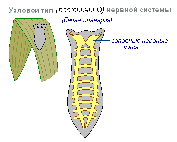
It is characterized by the fact that the connections of nerve cells are organized in a certain way, the excitation passes along strictly defined paths. This organization of the nervous system is more vulnerable. Damage to one node causes a violation of the functions of the whole organism as a whole, but it is faster and more accurate in its qualities.
tubular the nervous system is characteristic of chordates, it includes features of diffuse and nodular types. The nervous system of higher animals took all the best: high reliability of the diffuse type, accuracy, locality, speed of organization of reactions of the nodal type.
At the first stage of the development of the world of living beings, the interaction between the simplest organisms was carried out through the aquatic environment of the primitive ocean, into which chemical substances allocated by them. The first ancient form of interaction between the cells of a multicellular organism is chemical interaction through metabolic products entering the body fluids. Such products of metabolism, or metabolites, are the breakdown products of proteins, carbon dioxide, and others. This is the humoral transmission of influences, the humoral mechanism of correlation, or connections between organs.
The humoral connection is characterized by the following features:
Humoral connections are common to both the animal world and the plant world. At a certain stage in the development of the animal world, in connection with the emergence of the nervous system, a new, nervous form of connections and regulations is formed, which qualitatively distinguishes the animal world from the plant world. The higher the development of the animal organism, the greater the role played by the interaction of organs through the nervous system, which is designated as reflex. In higher living organisms, the nervous system regulates humoral connections. In contrast to the humoral connection, the nervous connection has an exact direction to a specific organ and even a group of cells; communication is carried out hundreds of times faster than the speed of distribution of chemicals. The transition from a humoral connection to a nervous one was accompanied not by the destruction of the humoral connection between the cells of the body, but by the subordination of nerve connections and the emergence of neurohumoral connections.
At the next stage in the development of living beings, special organs appear - glands, in which hormones are produced, which are formed from the nutrients entering the body. The main function of the nervous system is both in the regulation of the activity of individual organs among themselves, and in the interaction of the organism as a whole with its external environment. Any impact of the external environment on the body is primarily on the receptors (sense organs) and is carried out through changes caused by the external environment and the nervous system. As the nervous system develops, its highest department - the cerebral hemispheres - becomes "the manager and distributor of all the activities of the body."
The nervous system is made up of nervous tissue, which consists of a large number of neurons- a nerve cell with processes.

The nervous system is conditionally divided into central and peripheral.

central nervous system includes the brain and spinal cord, and peripheral nervous system- the nerves extending from them.

The brain and spinal cord are a collection of neurons. On the transverse section of the brain, white and gray matter are distinguished. The gray matter consists of nerve cells, and the white matter consists of nerve fibers, which are processes of nerve cells. In different parts of the central nervous system, the location of white and gray matter is not the same. In the spinal cord, gray matter is inside, and white is outside, while in the brain (cerebral hemispheres, cerebellum), on the contrary, gray matter is outside, white is inside. In different parts of the brain there are separate clusters of nerve cells (gray matter) located inside the white matter - nuclei. Accumulations of nerve cells are also located outside the central nervous system. They're called knots and belong to the peripheral nervous system.
The main form of activity of the nervous system is the reflex. Reflex- the reaction of the body to a change in the internal or external environment, carried out with the participation of the central nervous system in response to irritation of the receptors.
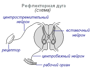
With any stimulation, excitation from the receptors is transmitted along the centripetal nerve fibers to the central nervous system, from where, through the intercalary neuron, along the centrifugal fibers, it goes to the periphery to one or another organ, the activity of which changes. This whole path through the central nervous system to the working organ is called reflex arc It is usually formed by three neurons: sensitive, intercalary and motor. A reflex is a complex act in which a much larger number of neurons take part. Excitation, getting into the central nervous system, spreads to many parts of the spinal cord and reaches the brain. As a result of the interaction of many neurons, the body responds to irritation.
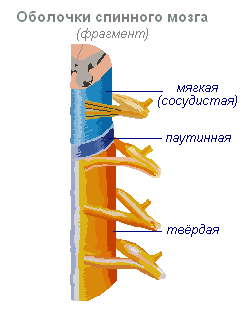
Spinal cord- a cord about 45 cm long, 1 cm in diameter, located in the spinal canal, covered with three meninges: hard, arachnoid and soft (vascular).
Spinal cord located in the spinal canal and is a strand, which at the top passes into the medulla oblongata, and at the bottom ends at the level of the second lumbar vertebra. The spinal cord is made up of gray matter containing nerve cells and white matter containing nerve fibers. Gray matter is located inside the spinal cord and is surrounded on all sides by white matter.

On a transverse section, the gray matter resembles the letter H. It distinguishes between the anterior and posterior horns, as well as the connecting crossbar, in the center of which there is a narrow spinal canal containing cerebrospinal fluid. Lateral horns are isolated in the thoracic region. They contain the bodies of neurons that innervate the internal organs. The white matter of the spinal cord is formed by nerve processes. Short processes connect parts of the spinal cord, and long ones make up the conductor apparatus of bilateral connections with the brain.
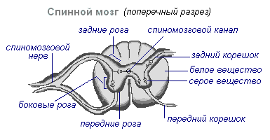
The spinal cord has two thickenings - cervical and lumbar, from which the nerves extend to the upper and lower extremities. There are 31 pairs of spinal nerves that emerge from the spinal cord. Each nerve starts from the spinal cord with two roots - anterior and posterior. back roots - sensitive composed of processes of centripetal neurons. Their bodies are located in the spinal nodes. Front roots - motor- are processes of centrifugal neurons located in the gray matter of the spinal cord. As a result of the fusion of the anterior and posterior roots, a mixed spinal nerve is formed. In the spinal cord centers are concentrated that regulate the simplest reflex acts. The main functions of the spinal cord are reflex activity and conduction of excitation.
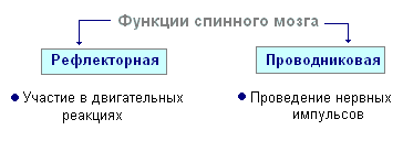
The human spinal cord contains the reflex centers of the muscles of the upper and lower extremities, sweating and urination. The function of conducting excitation is that impulses pass through the spinal cord from the brain to all areas of the body and vice versa. Centrifugal impulses from organs (skin, muscles) are transmitted to the brain along the ascending pathways. Centrifugal impulses are transmitted along descending paths from the brain to the spinal cord, then to the periphery, to the organs. If the pathways are damaged, there is a loss of sensitivity in various parts of the body, a violation of voluntary muscle contractions and the ability to move.
The formation of the central nervous system in the form of a neural tube first appears in chordates. At lower chordates neural tube persists throughout life higher- vertebrates - in the embryonic stage, the neural plate is laid on the dorsal side, which plunges under the skin and folds into a tube. In the embryonic stage of development, the neural tube forms three swellings in the anterior part - three cerebral vesicles, from which the brain regions develop: the anterior vesicle gives forebrain and diencephalon, middle vesicle turns into midbrain, posterior vesicle forms cerebellum and medulla oblongata. These five parts of the brain are characteristic of all vertebrates.
For lower vertebrates- fish and amphibians - the predominance of the midbrain over the rest of the departments is characteristic. At amphibians the forebrain is slightly enlarged and in the roof of the hemispheres thin layer nerve cells - the primary cerebral vault, the ancient cortex. At reptiles the forebrain is significantly enlarged due to accumulations of nerve cells. Most of the roof of the hemispheres is occupied by the ancient crust. For the first time in reptiles, the rudiment of a new bark appears. The hemispheres of the forebrain crawl onto other departments, as a result of which a bend is formed in the region of the diencephalon. Since the ancient reptiles, the cerebral hemispheres have become the largest part of the brain.
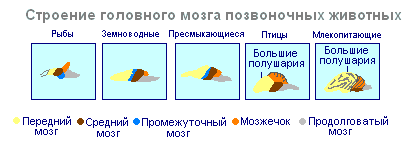
in the structure of the brain birds and reptiles much in common. On the roof of the brain is the primary cortex, the midbrain is well developed. However, in birds, compared with reptiles, the total mass of the brain and the relative size of the forebrain increase. The cerebellum is large and has a folded structure. At mammals the forebrain reaches its greatest size and complexity. Most of the medulla is the new cortex, which serves as the center of higher nervous activity. The intermediate and middle sections of the brain in mammals are small. The growing hemispheres of the forebrain cover them and crush them under them. In some mammals, the brain is smooth, without furrows and convolutions, but in most mammals there are furrows and convolutions in the cerebral cortex. The appearance of furrows and convolutions occurs due to the growth of the brain with a limited size of the skull. Further growth of the cortex leads to the appearance of folding in the form of furrows and convolutions.
If the spinal cord in all vertebrates is developed more or less equally, then the brain differs significantly in size and complexity of structure in different animals. The forebrain undergoes especially dramatic changes in the course of evolution. In lower vertebrates, the forebrain is poorly developed. In fish, it is represented by the olfactory lobes and nuclei of gray matter in the thickness of the brain. The intensive development of the forebrain is associated with the emergence of animals on land. It differentiates into the diencephalon and into two symmetrical hemispheres called telencephalon. Gray matter on the surface of the forebrain (cortex) first appears in reptiles, developing further in birds and especially in mammals. Indeed, large hemispheres of the forebrain become only in birds and mammals. In the latter, they cover almost all other parts of the brain.
The brain is located in the cranial cavity. It includes the brainstem and telencephalon (cerebral cortex).

brain stem consists of the medulla oblongata, pons, midbrain and diencephalon.
Medulla is a direct continuation of the spinal cord and expanding, passes into the hindbrain. It basically preserves the shape and structure of the spinal cord. In the thickness of the medulla oblongata are accumulations of gray matter - the nuclei of the cranial nerves. The rear axle includes cerebellum and pons. The cerebellum is located above the medulla oblongata and has a complex structure. On the surface of the cerebellar hemispheres, the gray matter forms the cortex, and inside the cerebellum, its nuclei. Like the spinal medulla oblongata, it performs two functions: reflex and conduction. However, the reflexes of the medulla oblongata are more complex. This is expressed in the importance in the regulation of cardiac activity, the state of blood vessels, respiration, sweating. The centers of all these functions are located in the medulla oblongata. Here are the centers of chewing, sucking, swallowing, separation of saliva and gastric juice. Despite its small size (2.5–3 cm), the medulla oblongata is a vital part of the CNS. Damage to it can cause death due to the cessation of breathing and heart activity. The conductive function of the medulla oblongata and the pons is to transmit impulses from the spinal cord to the brain and vice versa.
V midbrain primary (subcortical) centers of vision and hearing are located, which carry out reflex orientation reactions to light and sound stimuli. These reactions are expressed in various movements of the torso, head and eyes in the direction of stimuli. The midbrain consists of the cerebral peduncles and the quadrigemina. The midbrain regulates and distributes the tone (tension) of the skeletal muscles.
diencephalon consists of two departments - thalamus and hypothalamus, each of which consists of a large number of nuclei of the visual tubercles and the hypothalamic region. Through the visual hillocks centripetal impulses are transmitted to the cerebral cortex from all receptors of the body. Not a single centripetal impulse, no matter where it comes from, can pass to the cortex, bypassing the visual tubercles. Thus, through the diencephalon, all receptors are connected with the cerebral cortex. In the hypothalamic region there are centers that affect metabolism, thermoregulation and endocrine glands.
Cerebellum located behind the medulla oblongata. It is made up of gray and white matter. However, unlike the spinal cord and brainstem, the gray matter - the cortex - is located on the surface of the cerebellum, and the white matter is located inside, under the cortex. The cerebellum coordinates movements, makes them clear and smooth, plays an important role in maintaining the balance of the body in space, and also affects muscle tone. When the cerebellum is damaged, a person experiences a drop in muscle tone, movement disorder and a change in gait, speech slows down, etc. However, after some time, movements and muscle tone are restored due to the fact that intact parts of the central nervous system take over the functions of the cerebellum.
Large hemispheres- the largest and most developed part of the brain. In humans, they form the bulk of the brain and are covered with bark over their entire surface. Gray matter covers the outside of the hemispheres and forms the cerebral cortex. The cortex of the human hemispheres has a thickness of 2 to 4 mm and is composed of 6–8 layers formed by 14–16 billion cells, different in shape, size and functions. Under the bark is white matter. It consists of nerve fibers that connect the cortex with the lower sections of the central nervous system and the individual lobes of the hemispheres among themselves.
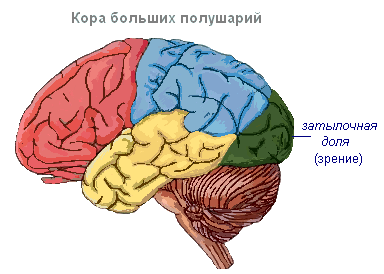
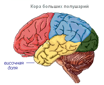
The cerebral cortex has convolutions separated by furrows, which significantly increase its surface. The three deepest furrows divide the hemispheres into lobes. There are four lobes in each hemisphere: frontal, parietal, temporal, occipital. Excitation of different receptors enters the corresponding perceiving areas of the cortex, called zones, and from here are transmitted to a specific organ, prompting it to action. The following zones are distinguished in the cortex. Hearing zone located in the temporal lobe, perceives impulses from auditory receptors.

visual area lies in the occipital region. This is where impulses come from the receptors of the eye.
Olfactory zone located on the inner surface of the temporal lobe and is associated with receptors in the nasal cavity.
Sensory-motor zone is located in the frontal and parietal lobes. In this zone are the main centers of movement of the legs, torso, arms, neck, tongue and lips. Here lies the center of speech.

The cerebral hemispheres are the highest division of the central nervous system that controls the functioning of all organs in mammals. The significance of the cerebral hemispheres in humans also lies in the fact that they represent the material basis of mental activity. I.P. Pavlov showed that physiological processes occurring in the cerebral cortex underlie mental activity. Thinking is connected with the activity of the entire cerebral cortex, and not only with the function of its individual areas.
| Department of the brain | Functions | |
| Medulla | Conductor | The connection between the spinal and overlying parts of the brain. |
| reflex | Regulation of the activity of the respiratory, cardiovascular, digestive systems:
|
|
| Pons | Conductor | Connects the hemispheres of the cerebellum to each other and to the cerebral cortex. |
| Cerebellum | Coordinating | Coordination of voluntary movements and maintaining the position of the body in space. Regulation of muscle tone and balance |
| midbrain | Conductor | Orienting reflexes to visual, sound stimuli ( head and body rotations). |
| reflex |
|
|
| diencephalon | thalamus
hypothalamus
|
|
Surface cerebral cortex in humans is about 1500 cm 2, which is many times greater than inner surface skulls. Such a large surface of the cortex was formed due to the development of a large number of furrows and convolutions, as a result of which most of the cortex (about 70%) is concentrated in the furrows. The largest furrows of the cerebral hemispheres - central, which runs across both hemispheres, and temporal separating the temporal lobe from the rest. The cerebral cortex, despite its small thickness (1.5–3 mm), has a very complex structure. It has six main layers, which differ in the structure, shape and size of neurons and connections. In the cortex there are centers of all sensitive (receptor) systems, representations of all organs and parts of the body. In this regard, centripetal nerve impulses from all internal organs or parts of the body approach the cortex, and it can control their work. Through the cerebral cortex, conditioned reflexes are closed, through which the body constantly, throughout life, very accurately adapts to the changing conditions of existence, to the environment.
The nervous system of higher animals and humans is the result of a long development in the process of adaptive evolution of living beings. The development of the central nervous system took place, first of all, in connection with the improvement in the perception and analysis of influences from the external environment. At the same time, the ability to respond to these influences with a coordinated, biologically expedient reaction was also improved. The development of the nervous system also proceeded in connection with the complication of the structure of organisms and the need to coordinate and regulate the work of internal organs.
The simplest unicellular organisms (amoeba) do not yet have a nervous system, and communication with the environment is carried out with the help of fluids that are inside and outside the body, - humoral or prenervous, form of regulation.
In the future, when the nervous system arises, another form of regulation appears - nervous. As it develops, it more and more subjugates the humoral, so that a single neurohumoral regulation with the leading role of the nervous system. The latter in the process of phylogenesis goes through a number of main stages.
Stage I - net nervous system. At this stage, the (intestinal) nervous system, such as hydra, consists of nerve cells, the numerous processes of which are connected to each other in different directions, forming a network that diffusely permeates the entire body of the animal. When any point of the body is stimulated, the excitation spreads throughout the entire nervous network and the animal reacts with the movement of the whole body. The diffuse nervous network is not divided into central and peripheral sections and can be localized in the ectoderm and endoderm.
Stage II - nodal nervous system. At this stage, (invertebrate) nerve cells converge into separate clusters or groups, and clusters of cell bodies produce nerve nodes - centers, and clusters of processes - nerve trunks - nerves. At the same time, the number of processes in each cell decreases and they receive a certain direction. According to the segmental structure of the body of an animal, for example, in an annelids, in each segment there are segmental nerve nodes and nerve trunks. The latter connect the nodes in two directions: the transverse shafts connect the nodes of a given segment, and the longitudinal ones connect the nodes of different segments. Due to this, nerve impulses that occur at any point in the body do not spread throughout the body, but spread along transverse trunks within this segment. Longitudinal trunks connect nerve segments into one whole. At the head end of the animal, which, when moving forward, comes into contact with various objects of the surrounding world, sensory organs develop, and therefore the head nodes develop more strongly than the others, giving rise to the development of the future brain. A reflection of this stage is the preservation of primitive features in humans (dispersion of nodes and microganglia on the periphery) in the structure of the autonomic nervous system.
Stage III - tubular nervous system. At the initial stage of animal development, a particularly important role was played by the apparatus of movement, on the perfection of which depended the main condition for the existence of an animal - nutrition (movement in search of food, capturing and absorbing it). In lower multicellular organisms, a peristaltic mode of locomotion has developed, which is associated with involuntary muscles and its local nervous apparatus. At a higher level, the peristaltic method is replaced by skeletal motility, i.e., movement with the help of a system of rigid levers - over the muscles (arthropods) and inside the muscles (vertebrates). The consequence of this was the formation of voluntary (skeletal) muscles and the central nervous system, which coordinates the movement of individual levers of the motor skeleton.
Such central nervous system in chordates (lancelet) it arose in the form of a metamerically built neural tube with segmental nerves extending from it to all segments of the body, including the apparatus of movement, the trunk brain. In vertebrates and humans, the trunk brain becomes the spinal cord. Thus, the appearance of the trunk brain is connected with the improvement, first of all, of the motor apparatus of the animal. The lancelet already has receptors (olfactory, light). The further development of the nervous system and the emergence of the brain is due mainly to the improvement of the receptor apparatus.
Since most of the sense organs arise at the end of the animal’s body that is turned in the direction of movement, i.e. forward, the anterior end of the trunk brain develops to perceive the external stimuli coming through them and the brain is formed, which coincides with the isolation of the anterior end of the body in the form of the head cephalization.
At the first stage development, the brain consists of three sections: posterior, middle and anterior, and from these sections in the first place (in lower fish) the posterior, or rhomboid brain, especially develops. The development of the hindbrain occurs under the influence of acoustic and gravity receptors (receptors of the VIII pair of cranial nerves, which are of leading importance for orientation in the aquatic environment). In the process of further evolution, the hindbrain differentiates into the medulla oblongata and the hindbrain proper, from which the cerebellum and pons develop.
In the process of adapting the body to the environment by changing the metabolism in the hindbrain, as the most developed section of the central nervous system at this stage, there are control centers for vital life processes associated, in particular, with the gill apparatus (respiration, blood circulation, digestion, etc.). .). Therefore, nuclei of the gill nerves arise in the medulla oblongata (group X of the pair - the vagus nerve). These vital centers of respiration and circulation remain in the human medulla oblongata. The development of the vestibular system associated with the semicircular canals and lateral line receptors, the emergence of nuclei of the vagus nerve and the respiratory center create the basis for the formation hindbrain.
At the second stage(still in fish) under the influence of the visual receptor, the midbrain especially develops. On the dorsal surface of the neural tube, a visual reflex center develops - the roof of the midbrain, where the fibers of the optic nerve come.
At the third stage, in connection with the final transition of animals from the aquatic environment to the air, the olfactory receptor is intensively developing, perceiving chemicals contained in the air, signaling prey, danger and other vital phenomena of the surrounding nature.
Under the influence of the olfactory receptor, the forebrain, prosencephalon, develops, initially having the character of a purely olfactory brain. In the future, the forebrain grows and differentiates into the intermediate and final. In the telencephalon, as in the higher part of the central nervous system, there appear centers for all kinds of sensitivity. However, the underlying centers do not disappear, but remain, obeying the centers of the overlying floor. Consequently, with each new stage in the development of the brain, new centers arise that subjugate the old ones. There is a kind of movement of functional centers to the head end and the simultaneous subordination of phylogenetically old rudiments to new ones. As a result, the centers of hearing that first appeared in the hindbrain are also present in the middle and forebrain, the centers of vision that arose in the middle are also present in the forebrain, and the centers of smell are only in the forebrain. Under the influence of the olfactory receptor, a small part of the forebrain, called the olfactory brain, develops, which is covered with a gray matter cortex - the old cortex.
The improvement of the receptors leads to the progressive development of the forebrain, which gradually becomes the organ that controls the entire behavior of the animal. There are two forms of animal behavior: instinctive, based on specific reactions (unconditioned reflexes), and individual, based on the experience of the individual (conditioned reflexes). According to these two forms of behavior, 2 groups of gray matter centers develop in the telencephalon: basal ganglia having the structure of nuclei (nuclear centers), and cortex of gray matter, which has the structure of a continuous screen (screen centers). In this case, the “subcortex” develops first, and then the cortex. The bark arises during the transition of an animal from an aquatic to a terrestrial lifestyle and is clearly found in amphibians and reptiles. The further evolution of the nervous system is characterized by the fact that the cerebral cortex more and more subjugates the functions of all underlying centers, there is a gradual function corticolization. The growth of the new cortex in mammals is so intense that the old and ancient cortex is pushed in the medial direction to the cerebral septum. The rapid growth of the crust is compensated by the formation of folding.
The necessary structure for the implementation of higher nervous activity is new bark, located on the surface of the hemispheres and acquiring a 6-layer structure in the process of phylogenesis. Due to the increased development of the new cortex, the telencephalon in higher vertebrates surpasses all other parts of the brain, covering them like a cloak. The developing new brain pushes the old brain (olfactory) into the depths, which, as it were, collapses, but remains as before the olfactory center. As a result, the cloak, that is, the new brain, sharply prevails over the rest of the brain - the old brain.
Rice. 1. Development of the telencephalon in vertebrates (according to Eddinger). I - human brain; II - rabbit; III - lizards; IV - sharks. Black indicates the new cortex, dotted line - the old olfactory part¸
So, the development of the brain takes place under the influence of the development of receptors, which explains the fact that the highest part of the brain: the brain - the cortex (gray matter) is a collection of cortical ends of the analyzers, that is, a continuous perceiving (receptor) surface.
The further development of the human brain is subject to other patterns associated with its social nature. In addition to the natural organs of the body, which are also found in animals, man began to use tools. Tools of labor, which became artificial organs, supplemented the natural organs of the body and constituted the technical "weapon" of man. With the help of this “weapon”, man acquired the opportunity not only to adapt himself to nature, as animals do, but also to adapt nature to his needs. Labor, as already noted, was a decisive factor in the formation of a person, and in the process of social labor, a means necessary for communication between people arose - speech. “First work, and then articulate speech along with it, were the two most important stimuli under the influence of which the brain of the monkey gradually turned into a human brain, which, for all its resemblance to the monkey, far surpasses it in size and perfection.” (K. Marx, F. Engels). This perfection is due to the maximum development of the telencephalon, especially its cortex - the new cortex.
In addition to analyzers that perceive various irritations of the outside world and constitute the material substrate of concrete-visual thinking characteristic of animals (the first signal system for reflecting reality, but to I.P. Pavlov), a person has the ability to abstract, abstract thinking with the help of a word, first heard ( oral speech) and later visible (written language). This constituted the second signaling system, according to I.P. Pavlov, which in the developing animal world was “an extraordinary addition to the mechanisms of nervous activity” (I.P. Pavlov). The material substrate of the second signal system became the surface layers of the new crust. Therefore, the cerebral cortex reaches its highest development in humans.
Thus, the evolution of the nervous system is reduced to the progressive development of the telencephalon, which in higher vertebrates and especially in humans, due to the complication of nervous functions, reaches enormous proportions. In the process of development, there is a tendency to move the leading integrative centers of the brain in the rostral direction from the midbrain and cerebellum to the forebrain. However, this trend cannot be absolutized, since the brain is an integral system in which stem parts play an important functional role at all stages of the phylogenetic development of vertebrates. In addition, starting from cyclostomes, projections of various sensory modalities are found in the forebrain, indicating the participation of this brain region in the control of behavior already at the early stages of vertebrate evolution.
Embryogenesis of the CNS.
Ontogenesis (ontogenesis; Greek op, ontos - existing + genesis - origin, origin) - the process of individual development of the organism from the moment of its inception (conception) to death. Allocate: embryonic (embryonic, or prenatal) - the time from fertilization to birth and postembryonic (post-embryonic, or postnatal) - from birth to death, periods of development.
The human nervous system develops from the ectoderm - the outer germ layer. At the end of the second week of embryonic development, a section of the epithelium separates in the dorsal parts of the body - neural (medullary) plate, cells of which intensively multiply and differentiate. The accelerated growth of the lateral sections of the neural plate leads to the fact that its edges first rise, then approach each other, and finally, at the end of the third week, grow together, forming the primary brain tube. After that, the brain tube gradually sinks into the mesoderm.
Fig.1. Formation of the neural tube.

The neural tube is the embryonic germ of the entire human nervous system. From it, the brain and spinal cord, as well as the peripheral parts of the nervous system, are subsequently formed. When the neural groove closes on the sides in the region of its raised edges (neural folds), a group of cells is isolated on each side, which, as the neural tube separates from the skin ectoderm, forms a continuous layer between the neural folds and ectoderm - the ganglionic plate. The latter serves source material for cells of sensitive nerve nodes (spinal and cranial ganglia) and nodes of the autonomic nervous system that innervates internal organs.
The neural tube at an early stage of its development consists of one layer of cylindrical cells, which subsequently intensively multiply by mitosis and their number increases; as a result, the wall of the neural tube thickens. At this stage of development, three layers can be distinguished in it: the inner one (later it will form the ependymal lining), the middle layer (the gray matter of the brain, the cellular elements of this layer differentiate in two directions: some of them turn into neurons, the other part into glial cells ) and the outer layer (white matter of the brain).
Fig.2. Stages of development of the human brain.

The neural tube develops unevenly. Due to the intensive development of its anterior part, the brain begins to form, cerebral bubbles form: first two bubbles appear, then the back bubble divides into two more. As a result, in four-week-old embryos, the brain consists of three brain bubbles(front, middle and rhomboid brain). At the fifth week, the anterior cerebral vesicle is subdivided into the telencephalon and diencephalon, and the rhomboid - into the posterior and medulla oblongata ( stage five brain bubbles). At the same time, the neural tube forms several bends in the sagittal plane.
The spinal cord with the spinal canal develops from the undifferentiated posterior part of the medullary tube. Formation occurs from the cavities of the embryonic brain brain ventricles. The cavity of the rhomboid brain is transformed into the IV ventricle, the cavity of the midbrain forms the aqueduct of the brain, the cavity of the diencephalon forms the III ventricle of the brain, and the cavity of the forebrain forms the lateral ventricles of the brain with a complex configuration.
After the formation of five cerebral vesicles in the structures of the nervous system, complex processes internal differentiation and growth of various parts of the brain. At 5-10 weeks, growth and differentiation of the telencephalon is observed: cortical and subcortical centers are formed, and the cortex is stratified. Meninges are formed. The spinal cord acquires a definitive state. At 10-20 weeks, the migration processes are completed, all the main parts of the brain are formed, and differentiation processes come to the fore. The end brain develops most actively. The cerebral hemispheres become the largest part of the nervous system. At the 4th month of human fetal development, a transverse fissure of the large brain appears, at the 6th - the central sulcus and other main sulci, in the following months - secondary and after birth - the smallest sulci.
In the process of development of the nervous system, myelination of nerve fibers plays an important role, as a result of which the nerve fibers are covered protective layer myelin and significantly increases the speed of nerve impulses. By the end of the 4th month of intrauterine development, myelin is detected in the nerve fibers that make up the ascending, or afferent (sensory) systems of the lateral cords of the spinal cord, while in the fibers of the descending, or efferent (motor) systems, myelin is found at the 6th month. At about the same time, myelination of the nerve fibers of the posterior cords occurs. Myelination of nerve fibers of the cortico-spinal tract begins in the last month of intrauterine life and continues for a year after birth. This indicates that the process of myelination of nerve fibers extends first to phylogenetically older structures and then to younger structures. The order of formation of their functions depends on the sequence of myelination of certain nerve structures. The formation of the function and also depends on the differentiation cellular elements and their gradual maturation, which lasts for the first decade.
By the time the baby is born, nerve cells reach maturity and are no longer capable of dividing. As a result, their number will not increase in the future. In the postnatal period, the final maturation of the entire nervous system gradually occurs, in particular its most complex section - the cerebral cortex, which plays a special role in the brain mechanisms of conditioned reflex activity, which is formed from the first days of life. Another important stage in ontogeny is the period of puberty, when sexual differentiation brain.
Throughout a person's life, the brain is actively changing, adapting to the conditions of the external and internal environment, some of these changes are genetically programmed, some are a relatively free reaction to the conditions of existence. The ontogenesis of the nervous system ends only with the death of a person.
Perm Institute of Humanities and Technology
Faculty of Humanities
TEST
in the discipline "ANATOMY OF THE CNS"
on the topic of
"The main stages of the evolutionary development of the central nervous system"
Perm, 2007
Stages of development of the central nervous system
The emergence of multicellular organisms was the primary stimulus for the differentiation of communication systems that ensure the integrity of the body's reactions, the interaction between its tissues and organs. This interaction can be carried out both in a humoral way through the entry of hormones and metabolic products into the blood, lymph and tissue fluid, and due to the function of the nervous system, which ensures the rapid transmission of excitation addressed to well-defined targets.
Nervous system of invertebrates
The nervous system as a specialized integration system on the path of structural and functional development passes through several stages, which in protostomes and deuterostomes can be characterized by features of parallelism and phylogenetic plasticity of choice.
Among invertebrates, the most primitive type of nervous system in the form diffuse neural network found in the intestinal type. Their nervous network is an accumulation of multipolar and bipolar neurons, the processes of which can cross, adjoin each other and lack functional differentiation into axons and dendrites. The diffuse nervous network is not divided into central and peripheral sections and can be localized in the ectoderm and endoderm.
epidermal nerve plexuses resembling the nervous networks of coelenterates can also be found in more highly organized invertebrates (flat and annelids), but here they occupy a subordinate position in relation to the central nervous system (CNS), which stands out as an independent department.
As an example of such centralization and concentration of nerve elements, one can cite orthogonal nervous system flatworms. The orthogon of higher turbellarians is an ordered structure, which consists of associative and motor cells, which together form several pairs of longitudinal cords, or trunks, connected by a large number of transverse and annular commissural trunks. The concentration of nerve elements is accompanied by their immersion into the depths of the body.
Flatworms are bilaterally symmetrical animals with a well-defined longitudinal body axis. Movement in free-living forms is carried out mainly towards the head end, where receptors are concentrated, signaling the approach of a source of irritation. These turbellarian receptors include pigment eyes, olfactory pits, statocysts, and sensory cells of the integument, the presence of which contributes to the concentration of nervous tissue at the anterior end of the body. This process leads to the formation head ganglion, which, according to the apt expression of Ch. Sherrington, can be considered as a ganglion superstructure over the systems of reception at a distance.
Ganglionization of nerve elements receives further development in higher invertebrates, annelids, mollusks and arthropods. In most annelids, the abdominal trunks are ganglionized in such a way that one pair of ganglia is formed in each segment of the body, connected by connectives to another pair located in the adjacent segment.
The ganglia of one segment in primitive annelids are interconnected by transverse commissures, and this leads to the formation ladder nervous system. In more advanced orders of annelids, there is a tendency for the abdominal trunks to converge up to the complete fusion of the ganglia of the right and left sides and the transition from the scalene to chain nervous system. An identical, chain type of structure of the nervous system also exists in arthropods with a different concentration of nerve elements, which can be carried out not only due to the fusion of neighboring ganglia of one segment, but also due to the fusion of successive ganglia of different segments.
The evolution of the nervous system of invertebrates goes not only along the path of concentration of nerve elements, but also in the direction of complication of structural relationships within the ganglia. It is no coincidence that modern literature notes the tendency to compare the ventral nerve cord with the spinal cord of vertebrates. As in the spinal cord, in the ganglia, a superficial arrangement of pathways is found, and the neuropil is differentiated into motor, sensory, and associative areas. This similarity, which is an example of parallelism in the evolution of tissue structures, does not, however, exclude the peculiarity of the anatomical organization. For example, the location of the trunk brain of annelids and arthropods on the ventral side of the body determined the localization of the motor neuropil on the dorsal side of the ganglion, and not on the ventral side, as is the case in vertebrates.
The process of ganglionization in invertebrates can lead to the formation scattered-nodular nervous system, found in molluscs. Within this numerous phylum there are phylogenetically primitive forms with a nervous system comparable to the orthogon of flatworms (lateral nerve molluscs) and advanced classes (cephalopods) in which fused ganglia form a differentiated brain.
The progressive development of the brain in cephalopods and insects creates a prerequisite for the emergence of a kind of hierarchy of command systems for controlling behavior. The lowest level of integration in the segmental ganglia of insects and in the subpharyngeal mass of the brain of mollusks, it serves as the basis for autonomous activity and coordination of elementary motor acts. At the same time, the brain is the following, a higher level of integration, where inter-analyzer synthesis and assessment of the biological significance of information can be carried out. On the basis of these processes, descending commands are formed that provide the variability in the launch of neurons of segmental centers. Obviously, the interaction of two levels of integration underlies the plasticity of the behavior of higher invertebrates, including innate and acquired reactions.
In general, when speaking about the evolution of the nervous system of invertebrates, it would be an oversimplification to represent it as linear process. The facts obtained in neurodevelopmental studies of invertebrates make it possible to assume a multiple (polygenetic) origin of the nervous tissue of invertebrates. Consequently, the evolution of the nervous system of invertebrates could proceed on a broad front from several sources with initial diversity.
At the early stages of phylogenetic development, a the second trunk of the evolutionary tree, which gave rise to echinoderms and chordates. The main criterion for distinguishing the type of chordates is the presence of a notochord, pharyngeal gill slits and a dorsal nerve cord - the neural tube, which is a derivative of the outer germ layer - the ectoderm. Tubular type of nervous system vertebrates, according to the basic principles of organization, is different from the ganglionic or nodal type of the nervous system of higher invertebrates.
Nervous system of vertebrates
Nervous system of vertebrates is laid in the form of a continuous neural tube, which in the process of ontogenesis and phylogenesis differentiates into various sections and is also a source of peripheral sympathetic and parasympathetic ganglions. In the most ancient chordates (non-cranial), the brain is absent and the neural tube is presented in an undifferentiated state.
According to the ideas of L. A. Orbeli, S. Herrick, A. I. Karamyan, this critical stage in the development of the central nervous system is designated as spinal. The neural tube of a modern non-cranial (lancelet), like the spinal cord of more highly organized vertebrates, has a metameric structure and consists of 62-64 segments, in the center of which passes spinal canal. The abdominal (motor) and dorsal (sensory) roots depart from each segment, which do not form mixed nerves, but go in the form of separate trunks. In the head and tail sections of the neural tube, giant Rode cells are localized, the thick axons of which form the conduction apparatus. The light-sensitive eyes of Hess are associated with Rode cells, the excitation of which causes negative phototaxis.
In the head part of the neural tube of the lancelet there are large ganglionic cells of Ovsyannikov, which have synaptic contacts with bipolar sensory cells of the olfactory fossa. Recently, neurosecretory cells resembling the pituitary system of higher vertebrates have been identified in the head of the neural tube. However, an analysis of the perception and simple forms of learning in the lancelet shows that at this stage of development the CNS functions according to the principle of equipotentiality, and the statement about the specificity of the head section of the neural tube does not have sufficient grounds.
In the course of further evolution, there is a shift of some functions and systems of integration from the spinal cord to the brain - encephalization process, which was considered on the example of invertebrates. During the period of phylogenetic development from the level of non-cranial to the level of cyclostomes the brain is formed as a superstructure over systems of distant reception.
A study of the central nervous system of modern cyclostomes shows that their rudimentary brain contains all the main structural elements. The development of the vestibulolateral system associated with the semicircular canals and lateral line receptors, the emergence of nuclei of the vagus nerve and the respiratory center create the basis for the formation hindbrain. The hindbrain of the lamprey includes the medulla oblongata and cerebellum in the form of small protrusions of the neural tube.
The development of distant visual reception gives impetus to laying midbrain. On the dorsal surface of the neural tube, the visual reflex center develops - the roof of the midbrain, where the fibers of the optic nerve come. And finally, the development of olfactory receptors contributes to the formation front or telencephalon, which is adjacent to the underdeveloped intermediate brain.
The direction of the encephalization process indicated above is consistent with the course of the ontogenetic development of the brain in cyclostomes. During embryogenesis, the head sections of the neural tube give rise to three cerebral vesicles. The telencephalon and diencephalon form from the anterior bladder, the middle bladder differentiates into the midbrain, and the medulla oblongata and cerebellum form from the posterior bladder. A similar plan of ontogenetic development of the brain is preserved in other classes of vertebrates.
Neurophysiological studies of the brain of cyclostomes show that its main integrative level is concentrated in the midbrain and medulla oblongata, i.e., at this stage of development, the central nervous system dominates bulbomesencephalic system of integration, replacing spinal.
For a long time, the forebrain of cyclostomes was considered purely olfactory. However, recent studies have shown that the olfactory inputs to the forebrain are not the only ones, but are complemented by sensory inputs from other modalities. Obviously, already at the early stages of vertebrate phylogenesis, the forebrain begins to participate in information processing and behavior control.
At the same time, encephalization as the main direction of brain development does not exclude evolutionary transformations in the spinal cord of cyclostomes. Unlike non-cranial neurons of skin sensitivity are isolated from the spinal cord and concentrated in the spinal ganglion. Improvement of the conductive part of the spinal cord is observed. The conductive fibers of the lateral columns have contacts with a powerful dendritic network of motor neurons. Downward connections of the brain with the spinal cord are formed through the Müllerian fibers - giant axons of cells lying in the midbrain and medulla oblongata.
The appearance of more complex forms of motor behavior in vertebrates, it is associated with the improvement of the organization of the spinal cord. For example, the transition from stereotypical undulating movements of cyclostomes to locomotion with the help of fins in cartilaginous fish (sharks, rays) is associated with the separation of cutaneous and musculo-articular (proprioceptive) sensitivity. Specialized neurons appear in the spinal ganglia to perform these functions.
Progressive transformations are also observed in the efferent part of the spinal cord of cartilaginous fishes. The path of motor axons inside the spinal cord is shortened, further differentiation of its pathways occurs. The ascending pathways of the lateral columns in cartilaginous fish reach the medulla oblongata and cerebellum. At the same time, the ascending pathways of the posterior columns of the spinal cord have not yet been differentiated and consist of short links.
The descending pathways of the spinal cord in cartilaginous fish are represented by a developed reticulospinal tract and pathways connecting the vestibulolateral system and the cerebellum with the spinal cord (vestibulospinal and cerebellospinal tracts).
At the same time, in the medulla oblongata there is a complication of the system of nuclei of the vestibulolateral zone. This process is associated with further differentiation of the lateral line organs and with the appearance in the labyrinth of the third (external) semicircular canal in addition to the anterior and posterior.
The development of general motor coordination in cartilaginous fish is associated with intensive development of the cerebellum. The massive cerebellum of the shark has bilateral connections with the spinal cord, medulla oblongata, and midbrain tegmentum. Functionally, it is divided into two parts: the old cerebellum (archicerebellum), associated with the vestibulo-lateral system, and the ancient cerebellum (fingerecerebellum), included in the proprioceptive sensitivity analysis system. An essential aspect of the structural organization of the cerebellum of cartilaginous fishes is its multi-layered nature. In the gray matter of the shark cerebellum, a molecular layer, a layer of Purkinje cells, and a granular layer were identified.
Another multilayer structure of the brainstem of cartilaginous fish is midbrain roof, where afferents of various modalities fit (visual, somatic). The very morphological organization of the midbrain indicates its important role in integrative processes at this level of phylogenetic development.
In the diencephalon of cartilaginous fish, differentiation of the hypothalamus, which is the most ancient formation of this part of the brain. The hypothalamus has connections with the telencephalon. The telencephalon itself grows and consists of olfactory bulbs and paired hemispheres. In the hemispheres of sharks, there are the rudiments of the old cortex (archicortex) and the ancient cortex (paleocortex).
The paleocortex, closely associated with the olfactory bulbs, serves mainly for the perception of olfactory stimuli. The archicortex, or hippocampal cortex, is designed for more complex processing of olfactory information. At the same time, electrophysiological studies have shown that olfactory projections occupy only part of the hemispheres of the forebrain in sharks. In addition to the olfactory, representation of the visual and somatic sensory systems was found here. Obviously, the old and ancient bark can participate in the regulation of search, feeding, sexual, and defensive reflexes in cartilaginous fishes, many of which are active predators.
Thus, in cartilaginous fishes, the main features of the ichthyopsid type of brain organization are formed. Its distinguishing feature is the presence of a suprasegmental integration apparatus that coordinates the work of motor centers and organizes behavior. These integrative functions are performed by the midbrain and cerebellum, which makes it possible to speak of mesenzphalocerebellar integration system at this stage of the phylogenetic development of the nervous system. The telencephalon remains predominantly olfactory, although it is involved in the regulation of the functions of the underlying sections.
The transition of vertebrates from an aquatic to a terrestrial way of life is associated with a number of rearrangements in the central nervous system. So, for example, in amphibians, two thickenings appear in the spinal cord, corresponding to the upper and lower limb girdle. In the spinal ganglia, instead of bipolar sensory neurons, unipolar neurons with a T-shaped branching process are concentrated, providing a higher rate of excitation without the participation of the cell body. On the periphery in the skin of amphibians are formed specialized receptors and receptor fields, providing discrimination sensitivity.
Structural changes also occur in the brain stem due to the redistribution of the functional significance of various departments. In the medulla oblongata, there is a reduction of the lateral line nuclei and the formation of a cochlear, auditory nucleus, which analyzes information from the primitive organ of hearing.
Compared to fish, amphibians, which have rather stereotyped locomotion, show a significant reduction in the cerebellum. The midbrain, like in fish, is a multilayered structure, in which, along with the anterior colliculus, the leading part of the integration of the visual analyzer, additional tubercles appear - precursors of the posterior colliculi of the quadrigemina.
The most significant evolutionary changes occur in the diencephalon of amphibians. Here is isolated visual tubercle - thalamus, structured nuclei appear (external geniculate body) and ascending pathways connecting the visual tubercle with the cortex (thalamocortical tract).
In the hemispheres of the forebrain, further differentiation of the old and ancient cortex occurs. In the old cortex (archicortex), stellate and pyramidal cells are found. In the gap between the old and ancient bark, a strip of a cloak appears, which is the forerunner new cortex (neocortex).
In general, the development of the forebrain creates the prerequisites for the transition from the cerebellar-mesencephalic integration system characteristic of fish to diencephalotelencephalic, where the forebrain becomes the leading department, and the visual tubercle of the diencephalon turns into a collector of all afferent signals. This integration system is fully represented in the sauropsid type of the brain in reptiles and marks the next stage of morphofunctional evolution of the brain .
The development of the thalamocortical system of connections in reptiles leads to the formation of new conducting pathways, as if pulling up to phylogenetically young brain formations.
In the lateral columns of the spinal cord of reptiles, an ascending spinothalamic tract, which conducts information about temperature and pain sensitivity to the brain. Here, in the side columns, a new descending tract is formed - rubrospinal(Monakova). It connects the motor neurons of the spinal cord with the red nucleus of the midbrain, which is included in the ancient extrapyramidal system of motor regulation. This multi-link system combines the influence of the forebrain, cerebellum, brainstem reticular formation, nuclei of the vestibular complex and coordinates motor activity.
In reptiles, as truly terrestrial animals, the role of visual and acoustic information increases, and it becomes necessary to compare this information with olfactory and gustatory information. Corresponding to these biological changes, a number of structural changes occur in the reptile brainstem. In the medulla oblongata, the auditory nuclei differentiate, in addition to the cochlear nucleus, an angular nucleus appears, connected with the midbrain. In the midbrain, the colliculus is transformed into the quadrigemina, in the posterior hills of which the acoustic centers are localized.
There is a further differentiation of connections between the roof of the midbrain and the thalamus, which is, as it were, the threshold to the entrance to the cortex of all ascending sensory pathways. In the thalamus itself, there is a further separation of nuclear structures and the establishment of specialized connections between them.
telencephalon reptiles can have two types of organization:
cortical and striatal. cortical organization type, characteristic of modern turtles, is characterized by the predominant development of the forebrain hemispheres and the parallel "development of new sections of the cerebellum. In the future, this direction in the evolution of the brain is preserved in mammals.
Striatal type of organization, characteristic of modern lizards, it is distinguished by the dominant development of the basal ganglia located in the depths of the hemispheres, in particular the striatum. This path is followed by the development of the brain in birds. It is of interest that in the striatum in birds there are cell associations or associations of neurons (from three to ten), separated by oligodendroglia. The neurons of such associations receive the same afferentation, and this makes them similar to neurons arranged in vertical columns in the neocortex of mammals. At the same time, identical cell associations have not been described in the striatum of mammals. Obviously, this is an example of convergent evolution, when similar formations developed independently in different animals.
In mammals, the development of the forebrain was accompanied by rapid growth of the neocortex, which is in close functional connection with the thalamus opticus of the diencephalon. Efferent pyramidal cells are laid in the cortex, sending their long axons to the motor neurons of the spinal cord.
Thus, along with the multilink extrapyramidal system, direct pyramidal pathways appear that provide direct control over motor acts. Cortical regulation of motor skills in mammals leads to the development of the phylogenetically youngest part of the cerebellum - the anterior part of the posterior lobes of the hemispheres, or neocerebellum. The neocerebellum acquires bilateral connections with the neocortex.
The growth of the new cortex in mammals is so intense that the old and ancient cortex is pushed in the medial direction to the cerebral septum. The rapid growth of the crust is compensated by the formation of folding. In the most poorly organized monotremes (platypus), the first two permanent furrows are laid on the surface of the hemisphere, while the rest of the surface remains smooth. (lissencephalic type of cortex).
As shown by neurophysiological studies, the brain of monotremes and marsupial mammals is devoid of the corpus callosum that still connects the hemispheres and is characterized by overlapping sensory projections in the new bark. There is no clear localization of motor, visual and auditory projections here.
Placental mammals (insectivores and rodents) develop a more distinct localization of projection zones in the cortex. Along with the projection zones, associative zones are formed in the neocortex, however, the boundaries of the first and second ones can overlap. The brain of insectivores and rodents is characterized by the presence of a corpus callosum and a further increase in the total area of the neocortex.
In the process of parallel-adaptive evolution, predatory mammals develop parietal and frontal associative fields, responsible for evaluating biologically significant information, motivating behavior and programming complex behavioral acts. Further development of folding of the new crust is observed.
Finally, primates show the highest level of organization of the cerebral cortex. The bark of primates is characterized by six layers, the absence of overlap of associative and projection zones. In primates, connections are formed between the frontal and parietal associative fields and, thus, an integral integrative system of the cerebral hemispheres arises.
In general, tracing the main stages of the evolution of the vertebrate brain, it should be noted that its development was not limited to a linear increase in size. In different evolutionary lines of vertebrates, independent processes of increasing the size and complication of the cytoarchitectonics of various parts of the brain could take place. An example of this is a comparison of the striatal and cortical types of organization of the vertebrate forebrain.
In the process of development, there is a tendency to move the leading integrative centers of the brain in the rostral direction from the midbrain and cerebellum to the forebrain. However, this trend cannot be absolutized, since the brain is an integral system in which stem parts play an important functional role at all stages of the phylogenetic development of vertebrates. In addition, starting from cyclostomes, projections of various sensory modalities are found in the forebrain, indicating the participation of this brain region in the control of behavior already at the early stages of vertebrate evolution.
Bibliographic list
1. Samusev R.P. Human anatomy. M., 1995.
2. Human anatomy. Ed. M.R. Sapina. M., 1986.
3. General course of human and animal physiology in 2 books. Ed. HELL. Nozdrachev. M., “Higher School”, 1991.


